Vasculitis refers to a group of inflammatory autoimmune diseases characterized pathologically by inflammation of the vascular walls. Systemic vasculitis involves multiple organs. Overall, systemic vasculitis has a low incidence and prevalence. Its clinical manifestations are complex and diverse, and it can result in irreversible damage to vital organs. Without treatment, mortality rates are high, making it a severe class of autoimmune diseases.
Nomenclature and Classification
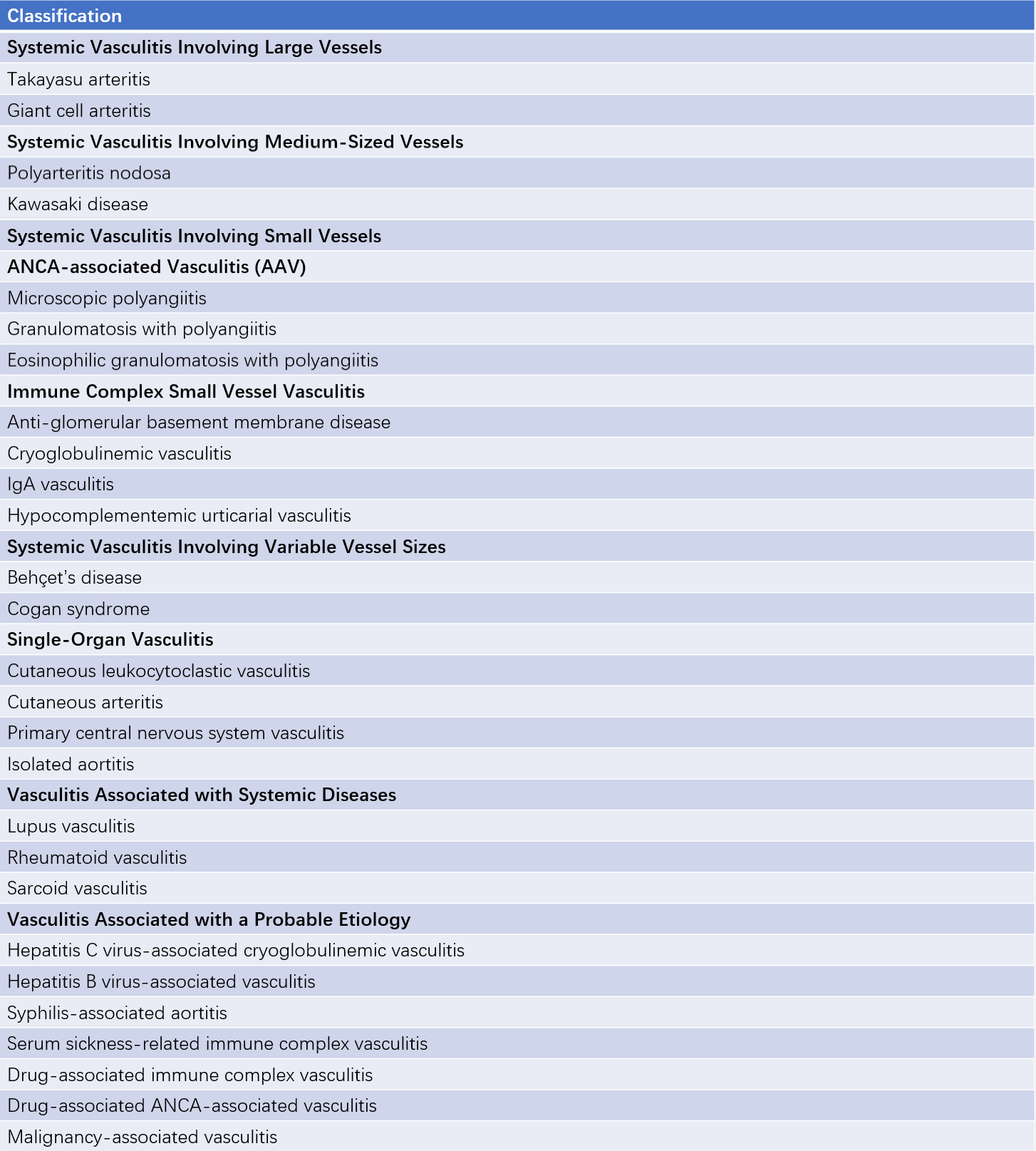
The currently accepted classification is based on the 2012 Chapel Hill Consensus Conference criteria, which categorizes vasculitis according to the size of the primarily affected blood vessels.

Table 1 2012 Chapel hill consensus conference classification of vasculitis
Etiology
The exact cause of vasculitis remains unclear. It is generally believed to be associated with genetic predisposition, infections, and environmental factors. Studies have identified genetic links such as HLA-DRB101 and HLA-DRB104 with susceptibility to giant cell arteritis, HLA-DRB52*01 with susceptibility to Takayasu arteritis, and HLA-DP/HLA-DQ genes with susceptibility to antineutrophil cytoplasmic antibody (ANCA)-associated vasculitis. Viral infections have also been implicated in vasculitis, as 10% of patients with polyarteritis nodosa (PAN) are associated with hepatitis B virus infection, and 80% of patients with mixed cryoglobulinemia are linked to hepatitis C virus infection. Mycobacterium tuberculosis infection has been associated with large-vessel vasculitis, such as Takayasu arteritis and Behçet's disease. Additionally, certain medications, including propylthiouracil, hydralazine, and cocaine, have been shown to trigger vasculitis by inducing the production of ANCA.
Pathogenesis
Although the precise mechanism remains unclear, it is thought to involve abnormalities in genetic susceptibility, infections, the innate immune system, and the adaptive immune system.
Infections
Direct vascular damage by external pathogens plays a role in disease initiation.
Macrophages and Their Cytokines
Some bacteria and viruses activate the innate immune system, including macrophages, through various pathways. Activated macrophages release pro-inflammatory cytokines such as tumor necrosis factor-α (TNF-α), interleukin-6 (IL-6), and IL-1, contributing to vascular wall inflammation.
Autoantibodies
Autoantibodies are critical in the development of vasculitis, with ANCA being the most significant. ANCA was the first autoantibody confirmed to contribute to vasculitis. Its target antigens include various components within the cytoplasm of neutrophils, such as proteinase 3 (PR3), myeloperoxidase (MPO), elastase, and lactoferrin, with PR3 and MPO being the primary target antigens.
The Complement System
The complement system, as part of the innate immune response, is also an essential component. In ANCA-associated vasculitis, neutrophils attacked by pathogens can activate the alternative complement pathway, releasing fragments such as C5a, leading to vascular damage and injury to tissues and organs.
Pathology
The fundamental pathological feature of vasculitis is inflammation and necrosis of the vascular wall. The key pathological changes include:
Vascular Wall Inflammation and Necrosis
These changes manifest as an infiltration of inflammatory cells, such as neutrophils, lymphocytes, and macrophages, as well as fibrinoid necrosis of the vascular wall. Fibrinoid necrosis is a characteristic pathological feature of vasculitis. In certain types of vasculitis, infiltrating inflammatory cells may also form multinucleated giant cells or granulomas composed of various inflammatory cells. For example, lymphocyte granulomas are present in granulomatosis with polyangiitis (GPA), while eosinophilic granulomas are seen in eosinophilic granulomatosis with polyangiitis (EGPA).
Destruction of Vascular Wall Structure
Inflammatory responses in the vascular wall can lead to collagen deposition, fibrosis, wall thickening, lumen narrowing, and secondary thrombus formation. Inflammation of the vascular wall can also result in damage to elastic fibers and smooth muscle, leading to aneurysm formation and vascular dilatation. In the same patient with vasculitis, multiple types of vascular lesions may coexist, and even within the same affected vessel, these pathological changes are often segmental.
Diagnosis
The diagnosis of vasculitis is challenging and requires a comprehensive assessment based on clinical manifestations, laboratory tests, pathological biopsies, and imaging studies to determine the type and extent of vascular involvement.
Clinical Manifestations
The clinical presentation of vasculitis mainly depends on the type and size of affected vessels and the involved organs, leading to diverse and nonspecific symptoms. Common manifestations include systemic symptoms caused by inflammation as well as organ-specific changes due to vascular inflammation, ischemia, and dysfunction. Systemic symptoms may include fatigue, fever, joint and muscle pain, and weight loss. Manifestations of organ involvement vary depending on the affected organ:
- Skin involvement may present as various types of rashes.
- Lung involvement may lead to cough, sputum production, hemoptysis, and dyspnea.
- Kidney involvement may cause proteinuria, hematuria, hypertension, and renal dysfunction.
- Nervous system involvement may result in headache, dizziness, altered consciousness, stroke, or clinical signs of peripheral neuropathy.
Diseases such as malignancies, infective endocarditis, fibromuscular dysplasia, atherosclerosis, and non-vasculitic embolic events (e.g., antiphospholipid syndrome, disseminated intravascular coagulation, cholesterol embolism, or tumor embolism) can mimic the clinical manifestations of systemic vasculitis and require careful differentiation.
Laboratory Testing
Most patients exhibit elevated white blood cell and platelet counts, chronic anemia, accelerated erythrocyte sedimentation rate (ESR), and increased C-reactive protein (CRP) during active disease. Kidneys may be affected, as indicated by hematuria, proteinuria, red blood cell casts, and elevated serum creatinine levels.
Special Investigations
ANCA (Antineutrophil Cytoplasmic Antibodies)
There are two methods for ANCA measurement, indirect immunofluorescence (IIF) and enzyme-linked immunosorbent assay (ELISA).
- IIF: Cytoplasmic fluorescence on neutrophils indicates C-ANCA positivity, while perinuclear fluorescence indicates P-ANCA positivity.
- ELISA: PR3-ANCA positivity is indicated by antibodies against proteinase 3 (PR3), and MPO-ANCA positivity is indicated by antibodies against myeloperoxidase (MPO).
ANCA is associated with small-vessel vasculitis. For example, C-ANCA is strongly associated with granulomatosis with polyangiitis (GPA), while P-ANCA is linked to microscopic polyangiitis (MPA) and eosinophilic granulomatosis with polyangiitis (EGPA). Together, GPA, MPA, and EGPA are collectively referred to as ANCA-associated vasculitides.
Anti-Endothelial Cell Antibody (AECA)
AECA has been identified in some cases of large-vessel vasculitis, such as Takayasu arteritis, Kawasaki disease, and Behçet’s disease, but it can also occur in various non-vasculitic conditions and infections. Its sensitivity and specificity for diagnosing vasculitis are relatively low.
Pathology
Biopsy represents the "gold standard" for diagnosing vasculitis. Pathological findings that support a diagnosis include inflammatory cell infiltration of the vascular walls, fibrinoid necrosis, granuloma formation, vascular lumen narrowing or occlusion, and thrombosis. Fibrinoid necrosis is considered a characteristic pathological change in vasculitis. However, as vascular lesions can be segmental, a biopsy showing no evidence of vascular inflammation does not definitively exclude vasculitis.
Angiography
Angiography serves as a key diagnostic tool for large- and medium-vessel vasculitis, providing the most definitive information on the extent of vascular lesions. Findings may include vascular wall thickening, narrowing, dilatation, aneurysm formation, and, in some cases, thrombosis.
Vascular Color Doppler Ultrasound
Doppler ultrasound is a non-invasive method suitable for evaluating the walls, lumens, and stenotic changes of larger and more superficial vessels. It is also useful for follow-up and comparative assessments over the course of the disease. However, its accuracy is lower than angiography and depends on the experience of the examiner.
CT Angiography (CTA)
CTA can visualize both the vascular walls and lumens as well as the extent of disease involvement. It can serve as an alternative to conventional angiography and is a key diagnostic tool for large- and medium-vessel vasculitis.
Magnetic Resonance Imaging (MRI)
Vascular MRI provides insights into the walls and lumens of large vessels and can evaluate active inflammation in the vascular wall. It holds significant value for diagnosing and assessing the disease status of large-vessel vasculitis.
Treatment Principles
Systemic vasculitis is generally progressive and can lead to irreversible organ damage without treatment. The principles of managing vasculitis include early diagnosis and early intervention. Treatment of systemic vasculitis is divided into two phases: induction of remission and maintenance of remission. The goal of the induction phase is to control acute vascular inflammation, maximize the recovery of organ function, and minimize irreversible organ damage. The objective of the maintenance phase is to keep the disease in a stable state, reduce the risk of relapse, minimize adverse effects from medications, and manage comorbid conditions.
Glucocorticoids are considered the cornerstone medication for treating vasculitis and represent the first-line treatment during the induction phase. Dosage and administration depend on the location and severity of the disease. For cases involving critical organs such as the kidneys, lungs, nervous system, or heart, higher doses of glucocorticoids are required, and the early addition of immunosuppressive agents is necessary.
Cyclophosphamide (CTX) is the most commonly used immunosuppressive agent, with well-established efficacy, although it is associated with significant adverse effects. Monitoring of blood counts, liver function, and other parameters is essential during therapy, and attention must be given to its gonadotoxicity. Other frequently used immunosuppressants include azathioprine, methotrexate, mycophenolate mofetil, and calcineurin inhibitors such as cyclosporine and tacrolimus.
In recent years, rituximab (RTX), a monoclonal antibody targeting CD20, has been successfully studied and approved for both induction and maintenance therapy in ANCA-associated vasculitides (AAV). For individuals with rapidly progressive kidney or lung involvement or critical illness, therapies such as plasma exchange, immunoadsorption, or high-dose intravenous immunoglobulin administration may be employed.