Systemic sclerosis (SSc), also known as scleroderma, is a complex autoimmune disorder characterized by autoimmunity, vascular abnormalities, and fibrosis. Clinically, it is distinguished by thickening and hardening of the skin and often affects multiple organs, including the cardiovascular system, lungs, digestive system, kidneys, and joints.
Globally, the annual incidence of SSc ranges from 8 to 56 new cases per 1 million people, with a prevalence of 38 to 341 cases per 1 million people. The majority of patients with SSc are women, with a female-to-male ratio of approximately 3–8:1. Although rare, SSc is one of the most fatal rheumatic diseases, associated with a significant disease burden.
Pathogenesis
The pathogenesis of SSc is complex and is believed to involve immune system dysfunction leading to damage and activation of vascular endothelial cells, which stimulate fibroblasts to produce excessive collagen, resulting in tissue fibrosis.
The development of SSc is influenced by multiple factors, including genetic predisposition, environmental factors, and sex hormones. Genetic studies have revealed associations with various HLA class II genes, and first-degree relatives of SSc patients are at a higher risk of developing SSc or other autoimmune diseases. Environmental factors play a significant role, as long-term exposure to substances such as polyvinyl chloride, organic solvents, epoxy resins, L-tryptophan, bleomycin, pentazocine, paclitaxel, and gemcitabine has been linked to skin hardening and visceral fibrosis. Higher disease prevalence has also been observed in individuals exposed to coal, gold mines, and silica dust, further emphasizing the role of environmental factors. Additionally, estrogen is thought to contribute to disease development, supported by the higher incidence of SSc in women compared to men, similar to other autoimmune diseases. Further studies are needed to investigate the interactions among these factors and the specific mechanisms underlying the disease.
Pathology and Pathophysiology
The fundamental pathological features of SSc include vascular changes and fibrosis caused by extracellular matrix overproduction.
Vascular Changes
Vascular abnormalities are observed in nearly all organs affected by SSc. Pathological findings include proliferative and occlusive vasculopathy, intimal hyperplasia of small- to medium-sized arteries, thickening of the basement membrane, and reduced capillary density accompanied by fibrosis. Perivascular inflammation is often seen in the early stages of the disease.
Fibrosis
Myofibroblasts produce excessive extracellular matrix components, resulting in edema and proliferation of collagen fibers. As the disease progresses, the edema subsides, and a marked increase in collagen fibers leads to tissue remodeling.
Clinical Manifestations
Subtypes
SSc can be classified into four subtypes:
Limited Cutaneous Systemic Sclerosis (lcSSc)
This subtype is characterized by skin involvement limited to areas distal to the elbows or knees, without extension beyond these joints, although the face and neck may also be affected. Disease progression is relatively slow, and patients often present with complications such as pulmonary arterial hypertension (PAH) and severe gastrointestinal involvement. Anti-centromere antibody (ACA) positivity is common in this subtype.
Diffuse Cutaneous Systemic Sclerosis (dcSSc)
This subtype involves more extensive skin involvement, including proximal limbs, the chest, and abdominal skin. The disease progresses rapidly and is associated with a poor prognosis due to frequent internal organ involvement. A high prevalence of anti-Scl-70 antibody positivity is noted. CREST syndrome, a specific variant of SSc, is characterized by calcinosis, Raynaud phenomenon, esophageal dysmotility, sclerodactyly, and telangiectasia. While primarily associated with lcSSc, it can also occur in dcSSc.
Systemic Sclerosis Without Skin Fibrosis (SSc Sine Scleroderma)
This rare form (affecting less than 5% of patients) is characterized by Raynaud phenomenon, distinctive internal organ manifestations (e.g., digital ulcers, PAH), and serological abnormalities, without clinically evident skin thickening.
Systemic Sclerosis Overlap Syndrome
Approximately 20% of systemic sclerosis (SSc) patients may have overlap with other connective tissue diseases, meeting the diagnostic criteria for two or more connective tissue disorders. These may include rheumatoid arthritis, systemic lupus erythematosus, or inflammatory myopathies, and this condition is referred to as systemic sclerosis overlap syndrome. This overlap syndrome can occur in any of the aforementioned SSc subtypes.
Symptoms
In addition to skin involvement, multi-organ involvement is a hallmark of the disease. Systematic assessment of organ involvement and regular evaluation of disease progression are critical for formulating personalized treatment plans and predicting prognosis.
Raynaud Phenomenon
Raynaud phenomenon is the most common initial symptom, occurring in over 90% of patients. It often emerges months to even decades before other manifestations of systemic sclerosis develop.
Skin
The skin is the most prominent organ affected, with involvement observed in over 95% of patients. Lesions typically begin in the fingers and toes, progressively spreading proximally in a symmetric distribution. Characteristic skin changes usually progress through three phases:
- Edematous Phase: The skin becomes swollen with non-pitting edema.
- Sclerotic Phase: The skin becomes firm and tight, resembling leather, and is difficult to pinch. Involvement of facial skin can erase normal facial wrinkles, resulting in a mask-like face with reduced facial expressions, a smaller and softer nose, thinner and retracting lips, perioral wrinkles, and a reduced mouth opening, which is referred to as a "mask facies."
- Atrophic Phase: If the disease remains uncontrolled, skin atrophy develops, making the skin smooth and thin and tightly adherent to the underlying bones. Joint contractures may also occur.
Skin involvement may exhibit pigmentation changes, with areas of hyperpigmentation interspersed with hypopigmentation, forming the so-called "salt-and-pepper sign." Digital ischemia can lead to loss of finger pad tissue, resulting in depressions, ulcers, and scars. Other features may include telangiectasia and subcutaneous calcification.
Lungs
High-resolution computed tomography (HRCT) reveals interstitial lung disease in 50–65% of patients, with non-specific interstitial pneumonia being the predominant histological pattern. Pulmonary arterial hypertension is another common pulmonary manifestation, particularly in patients with long-standing disease and extensive capillary damage. Uncontrolled progression may lead to right heart failure. The prognosis for SSc-related pulmonary hypertension is worse than that of idiopathic pulmonary arterial hypertension.
Heart
Cardiac involvement—which may include myocarditis, localized myocardial fibrosis, congestive heart failure, diastolic dysfunction, conduction abnormalities, and pericardial disease—is one of the leading causes of death in SSc. Most cases are asymptomatic and are detected only through investigations such as electrocardiography, echocardiography, or cardiac MRI.
Gastrointestinal Tract
More than 90% of patients exhibit gastrointestinal (GI) abnormalities. Esophageal involvement is the most commonly affected site, primarily involving dysfunction of the middle and lower esophagus. Capillary telangiectasia in the gastric antrum and intestines may lead to GI bleeding and anemia. Reduced gastrointestinal motility can cause abdominal distention, intestinal dilation, bacterial overgrowth, malabsorption syndrome, and pseudo-obstruction. Rectal dysfunction and anal sphincter damage may result in fecal incontinence. Nutritional issues associated with lower GI involvement require particular attention.
Kidneys
Renal tubular involvement is common. Renal crisis occurs in 1–14% of patients and manifests as acute renal failure and/or hypertensive emergencies, often accompanied by other microvascular abnormalities. Without timely intervention, renal failure may develop within weeks, resulting in significant mortality. Renal crisis is most frequently seen in early diffuse cutaneous SSc, particularly in individuals receiving high doses of corticosteroids or with positive anti-RNA polymerase III antibodies. Early use of angiotensin-converting enzyme inhibitors can significantly improve outcomes.
Joints, Bones, and Muscles
Fibrosis of the skin, fascia, and tendons surrounding joints can result in joint pain, reduced range of motion, and, in some cases, erosive arthritis. Late-stage disease may lead to joint contractures in abnormal fixed positions. Resorption of the finger bones may occur, leading to shortening of the digits.
Other Manifestations
Common non-specific symptoms include fatigue and depression. Ocular and oral dryness may occur. Neurological complications may include trigeminal neuralgia, carpal tunnel syndrome, and peripheral neuropathy. Male patients may develop erectile dysfunction. There is an increased risk of malignancies among patients with systemic sclerosis.
Physical Signs
Physical signs include Raynaud phenomenon, telangiectasia, and signs of skin involvement such as edema, tightness, atrophy, and ulcers. In classic cases, features such as "mask facies" and the "salt-and-pepper sign" are observable. Loss of soft tissue at the fingertips, leading to depressions, ulcers, and scarring, may also be present. Examination of other involved organs and tissues may reveal corresponding signs indicative of organ-specific pathology.
Auxiliary Examinations
Laboratory Tests
Autoantibodies assist in diagnosis and prognosis assessment. Up to 95% of patients test positive for antinuclear antibodies (ANA). Characteristic antibodies in SSc include anti-topoisomerase I (Scl-70) antibody, which is associated with diffuse cutaneous SSc, progressive pulmonary fibrosis, and digital ulcers; anticentromere antibody (ACA), which is commonly observed in limited cutaneous SSc and pulmonary arterial hypertension; and anti-RNA polymerase III antibody, which is linked to diffuse cutaneous SSc and renal crisis. Specific serum biomarkers may be elevated depending on organ involvement. For example, KL-6 levels are often elevated in patients with interstitial lung disease, while NT-proBNP levels tend to increase in cases of pulmonary arterial hypertension and cardiac involvement.
Imaging Studies
High-resolution computed tomography (HRCT) aids in the detection of interstitial lung disease and other SSc-associated abnormalities, such as esophageal dilation or pulmonary artery widening. Pulmonary function tests help in diagnosing interstitial lung disease.
Electrocardiography can identify cardiac arrhythmias. Transthoracic echocardiography is useful for screening pulmonary arterial hypertension, while right heart catheterization is required for confirmation. Early myocardial involvement can be detected through two-dimensional speckle tracking echocardiography. Cardiac MRI with late gadolinium enhancement suggests myocardial fibrosis. Patients with esophageal involvement may present with reduced or absent esophageal peristalsis, observed through barium swallow studies, which can also reveal dilation or stiffening of the esophagus. High-resolution esophageal manometry provides information on the presence and severity of esophageal motility disorders. Gastroscopy may reveal characteristic mucosal capillary telangiectasia in the stomach, appearing as wide linear streaks, known as "watermelon stomach."
Diagnosis
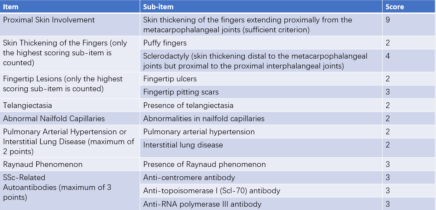
The diagnosis of SSc is based on Raynaud phenomenon, skin manifestations, characteristic visceral involvement, and detection of specific autoantibodies. Diagnostic criteria are guided by the 2013 American College of Rheumatology (ACR)/European League Against Rheumatism (EULAR) classification criteria and the 1980 American Rheumatism Association (ARA) classification criteria, which are more suitable for long-standing cases.

Table 1 2013 ACR/EULAR classification criteria for systemic sclerosis (SSc)
Differential Diagnosis
The differential diagnosis should exclude other diseases that cause skin thickening. Two exclusion criteria are particularly important:
- Skin thickening does not involve the fingers.
- Clinical features are more consistent with other conditions, such as nephrogenic systemic fibrosis, generalized morphea, eosinophilic fasciitis, diabetic sclerodema, scleromyxedema, erythromelalgia, porphyria, lichen sclerosus, graft-versus-host disease, and diabetic cheiroarthropathy.
Treatment
The primary goal of early treatment is to halt disease progression, whereas the aim of late-stage treatment is to improve quality of life and extend survival. Treatment plans should be individualized.
Immunosuppressants
For patients with severe or progressive disease, early initiation of immunosuppressive therapy is recommended. Commonly used agents include cyclophosphamide, mycophenolate mofetil, methotrexate, and tacrolimus. Biological agents such as rituximab and tocilizumab may be considered for refractory cases.
Antifibrotic Drugs
In rapidly progressing interstitial lung disease, antifibrotic drugs such as pirfenidone or nintedanib can be used in combination with other treatments.
Vasodilators
Vasodilatory agents are used to address vascular complications such as Raynaud phenomenon, digital ulcers, and pulmonary arterial hypertension. Depending on severity, medications may include calcium channel blockers, phosphodiesterase-5 (PDE-5) inhibitors, prostacyclin analogs, endothelin receptor antagonists, and soluble guanylate cyclase activators such as riociguat. Severe vascular conditions may require combinations of the aforementioned drugs. For scleroderma renal crisis, early initiation of angiotensin-converting enzyme inhibitors (ACEIs) or angiotensin receptor blockers (ARBs) is essential.
Glucocorticoids
Glucocorticoids show efficacy during the inflammatory phase for skin lesions, myositis, and interstitial lung disease. However, their use is associated with an increased risk of scleroderma renal crisis. It is generally advised to avoid high doses and to closely monitor blood pressure and renal function during use.
Cell-Based Therapies
Limited case reports and small-sample studies suggest that refractory patients may benefit from therapies such as autologous hematopoietic stem cell transplantation or CD19 CAR-T cell therapy. These approaches may be promising for patients with poor prognoses.
Other Therapeutic Measures
Patients with digital ulcers may require appropriate local wound care. Those with interstitial lung disease are encouraged to engage in tailored exercise programs to improve cardiopulmonary function. Proton pump inhibitors (PPIs) are effective in managing gastroesophageal reflux and preventing microaspiration.
Prognosis
The prognosis is poor, with death frequently resulting from involvement of the lungs, heart, kidneys, or gastrointestinal tract. Early diagnosis and the implementation of appropriate, individualized treatment are key to improving outcomes.