Atrophic rhinitis is a distinct type of rhinitis characterized by histopathological features such as atrophy or degenerative changes of the nasal mucosa. Primary atrophic rhinitis progresses slowly and has a long disease course. It is more commonly observed in females and individuals with frail constitutions. The disease is marked by nasal mucosal atrophy, reduced or lost sense of smell, and extensive crust formation within the nasal cavity. In severe cases, atrophy also extends to the periosteum and bony structures of the turbinates. The atrophic changes in mucosa can further extend to the nasopharynx, oropharynx, and laryngopharynx. In China, the incidence of this condition has shown a declining trend in recent years.
Etiology
The condition can be classified into primary and secondary types.
Primary Atrophic Rhinitis
The cause remains unknown. It is widely believed to result from a combination of internal and external factors. Contributing factors may include malnutrition, endocrine disorders, autonomic nervous system dysfunction, vitamin deficiencies (e.g., vitamins A, B, D, and E), genetic predisposition, low blood cholesterol levels, trace element imbalances or deficiencies, and immune dysregulation. Bacteria such as Klebsiella ozaenae and Corynebacterium diphtheriae-like organisms are not considered direct pathogens but may act as secondary infectious agents.
Secondary Atrophic Rhinitis
The condition may develop secondary to the following diseases or conditions:
- Chronic nasal or sinus infections with prolonged exposure to purulent secretions.
- Continuous exposure of the nasal mucosa to high concentrations of harmful dust or gases.
- Repeated or inappropriate nasal surgeries resulting in extensive damage to the mucosa (e.g., excessive resection of the inferior turbinates).
- Specific infectious diseases such as tuberculosis, syphilis, or leprosy that affect the nasal mucosa.
Pathology
The early phase shows only chronic inflammatory changes in the mucosa, which progress to atrophic degeneration. Pathological features include epithelial degeneration and atrophy, gradual development of obliterative arteritis and cavernous venous plexitis, proliferation and thickening of connective tissue in blood vessel walls, and narrowing or occlusion of blood vessel lumina. Poor vascular supply further contributes to the atrophy, fibrosis, and squamous metaplasia of the mucosa, glands, periosteum, and bone, along with fibrotic changes in the pterygopalatine ganglion.
Symptoms
Nasal obstruction
This may result from the accumulation of purulent crusts in the nasal cavity. Nasal obstruction can also occur despite unobstructed airflow, as mucosal sensory nerve atrophy and reduced sensitivity may lead to a subjective feeling of "nasal blockage.”
Dryness of the nose and throat
This occurs due to the atrophy of nasal mucosal glands, reduced secretions, or prolonged mouth breathing.
Epistaxis (nosebleeds)
Atrophic, thin, and dry nasal mucosa, as well as nose-picking or excessive nose-blowing, may lead to capillary rupture.
Reduced or lost sense of smell
This results from atrophy of the olfactory epithelium.
Foul odor
Decomposition of proteins in the crusts causes an offensive rotting odor during expiration, often referred to as “ozena.”
Headache or dizziness
These symptoms are linked to impaired nasal functions such as air-conditioning and humidification, as well as crust compression stimuli. The pain often involves the forehead, temple areas, or occipital region.
Examination
External Nose
Severe cases may result in deformities of the external nose, appearing as a flattened or saddle-shaped nose. Disease onset in childhood may affect normal nasal development.
Nasal Cavity Examination
The mucosa appears dry, the nasal cavity is abnormally wide, and the turbinates (especially the inferior turbinates) are markedly reduced in size. The nasal cavity contains a large quantity of crusts, which are yellow or greenish-yellow in color and emit a foul odor. If the pathology extends to adjacent areas such as the nasopharynx, oropharynx, or laryngopharynx, similar findings can be observed.
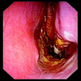
Figure 1 Atrophic rhinitis
Diagnosis and Differential Diagnosis
In severe cases, the characteristic symptoms and signs make diagnosis straightforward. Differential diagnosis should exclude specific nasal infectious diseases such as tuberculosis, syphilis, rhinoscleroma, nasal diphtheria, and nasal leprosy.
Milder cases may present with subtle signs like pale, thin, non-elastic mucosa (the turbinate appearing "bony") and a widened nasal cavity without significant crusting or noticeable olfactory loss.
Treatment
No definitive cure exists, and current management primarily involves local nasal irrigation and comprehensive systemic treatment.
Local Treatment
Nasal irrigation with physiological saline helps to clean the nasal cavity, remove crusts, and reduce foul odor.
Intranasal Medications
Compound menthol oil, liquid paraffin, or cod liver oil nasal drops are used to lubricate the mucosa, promote mucosal blood circulation, and soften crusts to facilitate their expulsion.
1% streptomycin nasal drops inhibit bacterial growth, reduce inflammatory ulceration, and support epithelial regeneration.
1% neostigmine mucosal application promotes vasodilation in nasal mucosa.
0.5% estradiol or hexestrol oil nasal drops reduce crust formation and mitigate foul odor.
50% glucose nasal drops may stimulate secretion by mucosal glands.
Surgical Treatment
Surgical intervention aims to reduce the size of the nasal cavity, thereby decreasing airflow, limiting evaporation of moisture from the nasal mucosa, and alleviating dryness and crust formation.
Systemic Treatment
Enhancing nutrition, improving environmental and personal hygiene, and supplementing vitamins are employed to protect the epithelial lining of the mucosa, enhance connective tissue's resistance to infection, promote cellular metabolism, dilate blood vessels, and improve circulation in the nasal mucosa. Supplementation with iron and zinc preparations may also have some therapeutic effects.
Complications
Complications of atrophic rhinitis include nasal dorsal collapse, nasal septal perforation, suppurative sinusitis, dacryocystitis, and secondary sinus mucoceles.