Atrioventricular block (AV block) occurs when the conduction of atrial impulses is delayed or blocked from reaching the ventricles after the atrioventricular junction exits its physiological refractory period. AV block can occur at different sites, including the AV node, His bundle, and bundle branches. It is classified into three degrees based on severity:
- First-degree AV block: Conduction time is prolonged, but all impulses still reach the ventricles.
- Second-degree AV block: It is divided into type I (Wenckebach) and type II.
Type I: Conduction time progressively lengthens until an impulse fails to reach the ventricles.
Type II: Intermittent conduction block, with two or more consecutive impulses failing to reach the ventricles, is often referred to as high-grade AV block.
- Third-degree AV block: It is also known as complete AV block, where no impulses are conducted to the ventricles, and the atria and ventricles are controlled by independent pacemakers.
Etiology
First-degree or second-degree type I AV block can occur in some healthy adults, children, and athletes, often transiently related to increased vagal tone at rest. Diseases causing AV block include acute myocardial infarction, coronary artery spasm, myocarditis/pericarditis, cardiomyopathy, acute rheumatic fever, cardiac tumors, congenital heart disease, myxedema, and infiltrative cardiac diseases (amyloidosis, sarcoidosis, or scleroderma). It can also result from electrolyte imbalances (hyperkalemia), drug toxicity (digitalis toxicity), and cardiac surgical injury (valve surgery, catheter ablation). In older adults, persistent AV block is often due to idiopathic degenerative changes in the conduction system, such as Lev's disease (calcification and sclerosis of the cardiac fibrous skeleton).
Clinical Manifestations
Patients with first-degree and second-degree type I AV block are usually asymptomatic. Second-degree type II AV block can cause skipped beats, leading to palpitations or perceived missed beats. High-grade AV block may present with dizziness, amaurosis fugax, or fatigue. Symptoms of third-degree AV block depend on ventricular rate and associated conditions, including fatigue, dizziness, syncope, angina, heart failure, and cerebral ischemia due to slow ventricular rate in severe patients, leading to transient loss of consciousness or seizures, known as Adams-Stokes syndrome, possibly resulting in sudden death.
On auscultation:
- First-degree AV block: Prolonged PR interval causes a weakened first heart sound.
- Second-degree type I AV block: Gradual weakening of the first heart sound is with skipped beats.
- Second-degree type II AV block: Intermittent skipped beats are with a constant first heart sound intensity.
- Third-degree AV block: AV dissociation leads to variable first heart sound intensity, normal or paradoxical splitting of the second heart sound, and occasionally loud accentuated first heart sounds (cannon sounds).
Electrocardiographic Features
First-degree Atrioventricular Block
The PR interval exceeds 0.20 seconds, with normal QRS complex morphology and duration.

Figure 1 First-degree AV block, PR interval = 0.37 seconds
Second-degree Atrioventricular Block
It is divided into type I and type II. Type I, also known as Wenckebach block, is the most common form of second-degree AV block.
Type I (Wenckebach) Block
Regular P waves are with progressively lengthening PR intervals until a P wave is blocked, resulting in a dropped QRS complex. Common AV conduction ratios are 3:2 and 5:4. The block is usually at the AV node, with normal QRS complexes. This type rarely progresses to third-degree AV block.

Figure 2 Second-degree Type I AV block
In lead II, P waves appear regularly. The 3rd and 7th P waves from the left (indicated by arrows) are not conducted to the ventricles, showing 3:2 and 4:3 AV conduction.
Type II Block
Constant PR interval with some P waves is not followed by QRS complexes. If the QRS complex is normal, the block may be at the AV node. If the QRS complex is widened and abnormal, the block is in the His-Purkinje system.

Figure 3 Second-degree Type II AV Block
In lead II, P waves appear regularly. The 3rd and 6th P waves from the left (indicated by arrows) are not conducted to the ventricles, showing 3:2 AV conduction.
High-grade AV Block
Two or more consecutive P waves fail to conduct to the ventricles, often with 2:1 or 3:1 AV conduction.

Figure 4 High-grade AV block with junctional escape
In leads II and III, P waves appear regularly. The 2nd and 3rd P waves from the left (indicated by arrows) are not conducted to the ventricles, while the 4th P wave is conducted, showing 3:1 AV conduction.
Third-degree (Complete) Atrioventricular Block
ECG features include:
- Independent rhythms of P waves and QRS complexes
- Atrial rate faster than ventricular rate, with atrial impulses originating from the sinus node or an ectopic atrial rhythm (atrial tachycardia, flutter, or fibrillation)
- Ventricular pacemaker usually just below the block site

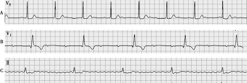
Figure 5 Third-degree AV block
A. In lead V5, P waves have a slightly irregular rhythm, with an average atrial rate of 75 bpm. The QRS complexes are normal in morphology and regular, with a ventricular rate of 50 bpm.
B. In lead V1, P waves have a regular rhythm, with an atrial rate of 83 bpm. The QRS complexes are widened and regular, with a ventricular rate of 29 bpm.
C. In lead II, P waves are absent and replaced by f waves of varying size, shape, and intervals, accompanied by delayed and widened QRS complexes. The RR intervals are roughly equal, with a ventricular rate of 30 bpm.
If the pacemaker is at the His bundle or above, ventricular rate is 40 - 60 bpm, with normal QRS complexes and relatively stable rhythm; if below the His bundle, ventricular rate may be below 40 bpm, with widened QRS complexes and often unstable ventricular rhythm.
Treatment
Treatment should first address the underlying cause. First-degree and second-degree type I AV blocks generally require no specific treatment. For symptomatic patients with slow ventricular rates, temporary or permanent cardiac pacing should be initiated early. Irreversible second-degree type II, high-grade, and third-degree AV blocks require permanent pacing regardless of symptoms. During periods without pacing options or while awaiting pacing, atropine (0.5 - 2.0 mg IV) can be used to increase heart rate in AV block; isoproterenol (1 - 4 μg/min IV infusion) is suitable for any cardiac conduction block but should be used cautiously or avoided in acute myocardial infarction. Prolonged use of these drugs often yields poor results and can cause severe adverse effects, so they are only suitable for emergencies without pacing options.